Computed tomography is a relatively new (invented in 1972) and highly informative method of medical diagnostics, which is based on X-rays. The device is designed in such a way that the X-ray tube and many rows of sensors rotate around the area under study and take many “frames” at once, which are then transferred to a computer, which processes all the received images and builds sections from them …
Everything you need to know about CT
Articles 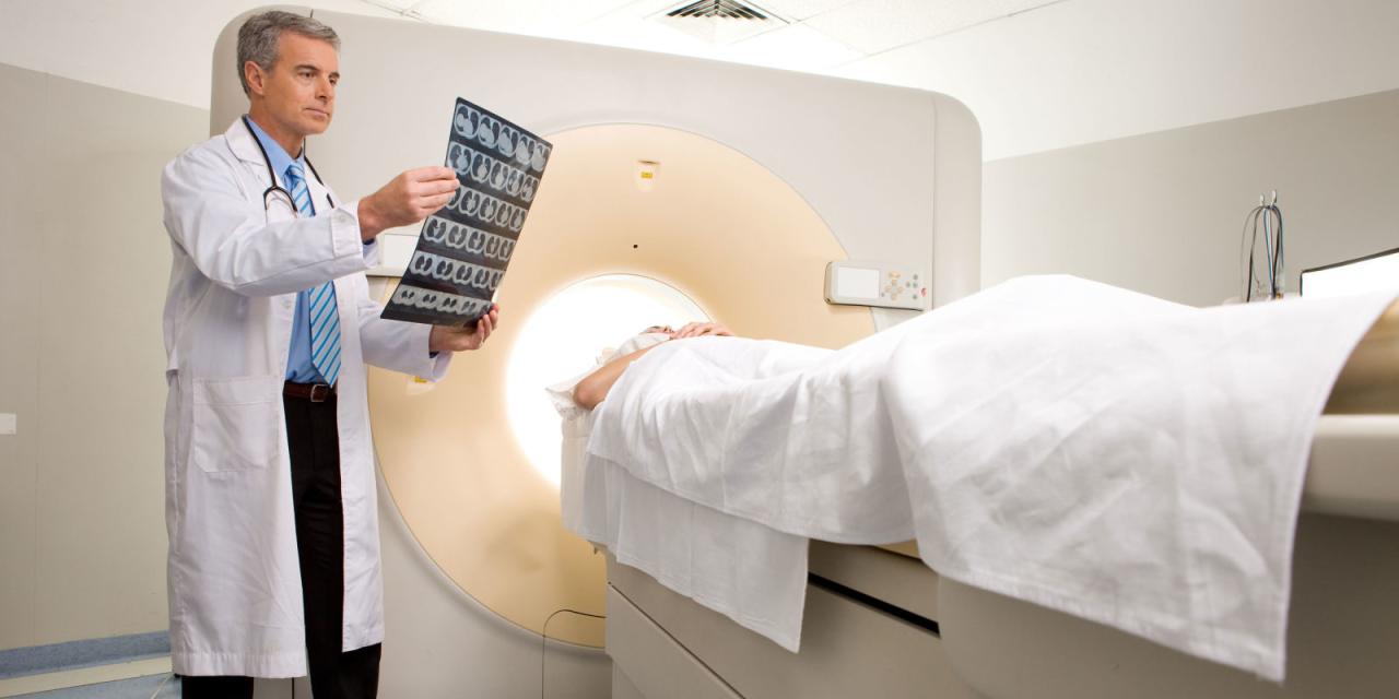
Computed tomography is a relatively new (invented in 1972) and highly informative method of medical diagnostics, which is based on X-rays.
The device is designed in such a way that the X-ray tube and many rows of sensors rotate around the area under study and take many “frames” at once, which are then transmitted to a computer, which processes all the received images and builds sections of the human body on the monitor from them.
Computed tomography is completely painless and does not cause claustrophobia in the patient, the patient remains dressed. The scanning procedure takes less than one minute. In the event that the examination is carried out using contrast enhancement, the scan is repeated several times. At the end of the procedure, the radiologist examines the received images on the computer screen and writes a conclusion.
How is a CT scan performed?
The X-ray laboratory assistant performs a special laying of the patient, depending on the examination. Further, the gantry (the device on which the tube and receiving sensors are installed) begins to rotate around the scanned area and makes slices of the area under study with certain parameters of power and the tube passage step. When scanning the organs of the chest, abdominal cavity, you will need to hold your breath several times, about which the X-ray laboratory assistant will warn you. The scanning procedure lasts 2-4 minutes.
What "dislikes" SKT?
First of all, it's movement. Any movements during scanning of the study area distort the resulting images, of course, and reduce their information content for the doctor. If even only 1-2 scans in a series of obtained images are distorted by motion artifacts, then subsequent 3D reconstructions will already be defective and their information content for the doctor, of course, will be reduced.
Secondly, metal. Metal is to X-rays like a mountain to the sun – it leaves a "shadow". A large amount of metal in the study area can significantly distort the resulting images. But if the metal is located in any other place, except for the area that is being examined, it will not be able to affect the quality of the scans in any way.
What do I need to bring with me for a CT scan?
Before the examination, it is necessary to give the X-ray laboratory assistant or radiologist the medical documentation you have (a referral from the attending physician, which indicates the preliminary diagnosis,Research zone, clinical task, specific comments), data of previous studies (which directly concern the cause of the assignment to you) and other medical records, analyzing the radiologist to formulate the most accurate diagnostic conclusion on CT, taking into account the data of your medical card and the recommendations of the attending Doctor. If you do not provide medical documentation in full, then the radiologist assesses only directly obtained KT scans, without taking into account previous injuries and diseases, all sorts of consequences that they could entail and exclude possible concomitant pathology.
Remember, there is no absolute and universal method of medical diagnosis: MRI will never replace CT, CT will never replace ultrasound, ultrasound never replace radiography. All these methods complement each other and sometimes, the data of previous studies have a decisive word in the correct interpretation of the resulting picture by a radiologist during computed tomography.
Who decrypts the results and when can I get them?
The obtained images of the CT should explore the radiologist (a doctor who specializes in CT, MRI, X-ray studies having an appropriate certificate). It is necessary to "read" all scans, explore the provided honey. Documentation, compare it with the resulting CT-picture, describe the study, withdraw a diagnostic conclusion, to extract the film with scannes. On average, it takes about 1-3 hours. In the Mediskan diagnostic center, we undertake to prepare results for extradition on the next business day.
After the study, you get films with scans and diagnostic conclusion, which will be signed by a radiologist. Also in diagnostic conclusion there may be recommendations for your further actions (to which doctor to handle these results, which types of finishing are recommended and so on).
Is computer tomography safe?
Computer tomography is considered a secure method. The dose of X-ray irradiation is relatively small. Modern technologies allowed to reduce the dose of irradiation to a minimum. By radial load, it can be compared with the load when flying by plane. Dose of irradiation depends on the size of the scanned area. Accordingly, when scanning an abdominal cavity, the dose of irradiation is higher than when scanning the foot. There is also a very small risk if the introduction of sedatives and contrast agents is required. The patient must warn a doctor if he has allergies for medicines, iodine, seafood, if he suffers with diabetes, asthma, heart disease and thyroid gland.
What is the difference between CT and MRI?
Computer tomography (CT) is a radiation diagnostic method based on the use of X-ray rays.A CT scanner is a special X-ray machine that rotates around the patient's body and takes pictures from various angles. The images are processed and summarized by a computer. Computed tomography is based on the measurement and complex computer processing of how much X-ray radiation is attenuated in tissues of various density.
Magnetic resonance imaging (MRI) does not use ionizing radiation, but uses the action of magnetic fields. To obtain an image, the patient's magnetic resonance imaging (MRI) scanner is placed in a strong magnetic field, which causes all the hydrogen atoms in the patient's body to line up parallel to the direction of the magnetic field. At this moment, the device sends an electromagnetic signal, perpendicular to the main magnetic field. Hydrogen atoms, which have the same frequency as the signal, are “excited” and generate their own signal, which is picked up by the apparatus. Different types of tissues (bones, muscles, blood vessels, etc.) have a different number of hydrogen atoms and therefore they generate a signal with different characteristics. The tomograph recognizes these signals, decodes them and builds an image. Thus, tissues are differentiated by the content of hydrogen atoms, to put it simply, water.
That is why magnetic resonance imaging has its own contraindications, including the first trimester of pregnancy, the presence of a pacemaker, middle ear implants, metal foreign bodies.
What are the contraindications for CT?
The main contraindication for CT scan is pregnancy and patient weight over 150 kg. If the study is performed with contrast, you should make sure that the patient is not allergic to the iodine-containing contrast agent. In case of renal or hepatic insufficiency, it is necessary to consult a radiologist before the examination.
How often can a CT be done?
There are no strict restrictions on the number of CT procedures for a certain period, however, it is desirable to take into account the total radiation exposure from studies conducted during the year.
Can children do CT?
For children under 14 years of age, computed tomography is performed only when indicated. Since the study is associated with radiation exposure, when examining young children, it is necessary to carefully weigh the need for MSCT in each case.
Why is pregnancy a contraindication for CT?
CT is not performed on pregnant women, as it is an X-ray method of examination. Exceptions are cases associated with a threat to life. MSCT is especially contraindicated in the first trimester of pregnancy. For diagnosis in pregnant women, other methods are used: ultrasound diagnostics and magnetic resonance imaging. In rare cases, when MSCT is indispensable (for example, in severe trauma), it is still done, but at the same time the uterus is covered with a lead screen.If you are pregnant, be sure to report this to the doctor conducting tomography.
Is it possible to pass CT during lactation?
If the CT was carried out without using a contrast agent, then breastfeeding can be breastfeed directly after the procedure. When applying contrast, it does not recommend breastfeeding within 48 hours after the study.
What time does computed tomography take?
Scanning procedure takes less than a minute. In the event that the study is carried out using contrast gain, the scanning is repeated several times, and the entire procedure lasts up to an hour.
Is the irradiation harmful, which I get during the study?
The exposure that you get in the study on CT is similar to irradiation in an ordinary x-ray study. You are also subjected to a small effects of radiation. But the doctors and other scientists believe that this risk is quite acquitted.