Vesicoureteral reflux is a condition characterized by the backflow of urine from the bladder into the ureter. Occurs with abnormalities of the excretory system, high pressure inside the bladder or against the background of inflammatory processes. Reflux can cause pyelonephritis, hydronephrosis, and kidney failure. The main symptoms are pain in the lumbar region after urination, cloudy urine, swelling, and fever. Diagnostic methods: general urine and blood tests, ultrasound of the kidneys, excretory urography, voiding cystography. Treatment is reduced to therapy of an inflammatory disease or surgical elimination of anomalies of the urinary system.
Vesicoureteral reflux (Vesicoureteral reflux)

Vesicoureteral reflux is a pathology characterized by the reverse flow of urine from the bladder into the ureter. Occurs with abnormalities of the excretory system, high pressure inside the bladder or against the background of inflammatory processes. Reflux can cause pyelonephritis, hydronephrosis, and kidney failure. The main symptoms are pain in the lumbar region after urination, cloudy urine, swelling, and fever. Diagnostic methods: general urine and blood tests, ultrasound of the kidneys, excretory urography, voiding cystography. Treatment is reduced to the treatment of an inflammatory disease or the surgical removal of anomalies of the urinary system.
SME-10.

General
Vesicoureteral or vesicoureteral reflux is one of the most common urological diseases, especially among children. It is found in 1% of urological patients, the proportion of the bilateral process is 50.9%. Urinary regurgitation is detected in 40% of patients with urinary tract infections.
The prevalence of pathology, a high risk of complications (renal failure, secondary arterial hypertension, purulent kidney disease) determine a large percentage of patients with disability. Congenital reflux occurs in 1 in 100 children, with a female to male ratio in the first year of life of 5:1. As they grow older, the frequency of occurrence of pathology in boys increases with a change in the situation to the opposite.

Causes
The etiological factors of non-physiological movement of urine are processes leading to insufficiency of the sphincter of the ureteral anastomosis. The sphincter is a physiological barrier that separates the ureters from the bladder and prevents backflow of urine. Additional prerequisites for regurgitation creates a high fluid pressure in the bladder. The main groups of factors leading to the development of reflux include:
- Anomalies in the development of the excretory system . A decrease in the closing function of the sphincter develops due to the incorrect formation of this segment of the excretory system at the stage of intrauterine development. The abnormal structure can manifest itself in the form of a permanently open ureteral orifice, the absence or reduction of the muscle layer of the constrictor, its dysplasia, and tissue degeneration.
- High intravesical pressure of urine . Damage to the brain, spinal cord, pelvic nerves leads to dysregulation of bladder muscle tone. The muscle wall is in constant tension, which creates increased hydrostatic pressure. This results in the inability of the healthy sphincter to hold back urine. The causative factors of this condition are congenital (infantile cerebral palsy, sacral agenesis) and acquired (brain tumors, stroke, Parkinson’s disease, diabetes mellitus) pathologies.
- Inflammatory process . A decrease in the barrier function of the vesicoureteral fistula is possible with inflammation of the urinary tract. Reflux is usually the result of advanced acute and chronic forms of cystitis or ascending urethritis. The infection is more often caused by opportunistic microorganisms, especially E. coli, against the background of a decrease in local or general immunity.
- iatrogenic causes . The formation of retrograde reflux of urine through the vesicoureteral fistula is possible after surgery in the area of the distal excretory apparatus. The most frequent operations leading to reflux are prostatectomy, dissection of the ureterocele, resection of the bladder neck. With any of them, there is a possibility of a violation of the normal anatomical structure of the bladder and vesicoureteral segment.
Factors that increase the risk of developing reflux include its presence in the family history, especially in the immediate family (parents, brothers, sisters). Also increase the likelihood of violations of the regulation of the tone of the bladder or the sphincter of the anastomosis of the tumor of the spinal cord, congenital anomalies of the spine, for example, its splitting.
Pathogenesis
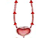
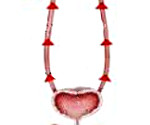
The area of connection of the ureters with the cavity of the bladder anatomically represents a sphincter antireflux apparatus, which ensures the flow of urine only in a downward direction. This is achieved due to a certain angle at which the ureter flows into the bladder, and intramural smooth circular muscles. The main pathological link in the formation of reflux is a decrease in the efficiency of the sphincter as a result of dysplasia of muscle fibers, their inflammatory damage, and impaired nervous regulation. Morphofunctional changes lead to disruption of the antireflux mechanism and non-physiological retrograde movement of urine.
High hydrostatic pressure causes deformation and dilatation of the ureter and renal pelvis. Conditions are created for the transfer of bacteria from the lower segments of the excretory system to the upper ones, which leads to the development of an acute or chronic recurrent infection in the kidney parenchyma with the replacement of renal tissue with non-functional connective tissue. Nephrosclerosis is the cause of renal filter dysfunction and the development of life-threatening conditions.
Classification
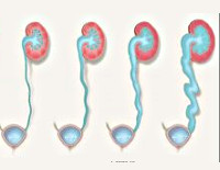
Modern clinical urology strives to develop a unified generally recognized classification, since the choice of further therapeutic tactics largely depends on the degree of vesicoureteral reflux (VUR). To date, the most widespread systematization of the process, depending on the level of urine reflux:
- I degree . Due to insufficiency of the sphincter, the reflux of a small amount of urine is limited to the distal pelvic ureter. There is no dilation of the ureter. The risk of complications of an infectious and non-infectious nature is minimal, and there are no symptoms. Detection of VUR usually occurs during examination for other diseases of the excretory system.
- II. degree . Urine reflux is noted throughout the ureter, but without its dilatation. In this case, urine does not reach the kidneys, pyelocaliceal system. This degree is characterized by the absence of pronounced symptoms, a small risk of infectious complications, but a high rate of progression of reflux, a rapid transition to the next levels of development. It is discovered by chance during a routine preventive examination or diagnosis of other pathologies.
- III degree . Urine reaches the kidneys, but there is no expansion of the pelvis. Perhaps a decrease in renal function by 20%, found in biochemical analyzes. The ureter is dilated, there are signs of degenerative trophic degeneration of tissues. The risk of infection is increased due to stagnation of urine in the excretory system, which is often the reason for contacting a specialist. Symptoms are moderate.
- IV degree . A significant expansion, deformation of the pyelocaliceal region and ureters is recorded. Kidney function decreases significantly (up to 50%) with a decrease in urine production, especially against the background of infectious complications. Symptoms are pronounced, with febrile temperature, generalized edema. With a bilateral process, the development of life-threatening conditions is possible, which requires an early appeal to specialists.
- V degree . A severe degree of kidney damage is diagnosed with thinning of their parenchyma, along with all the signs characteristic of previous degrees. The ureter, due to excessive expansion, has knee-shaped bends. Increasing symptoms of renal failure (decreased diuresis, nausea, vomiting, pruritus) require immediate treatment for qualified help.
There are classifications of vesicoureteral reflux based on other signs, for example, on the etiological factor (congenital, acquired), the nature of the process (unilateral, bilateral), and the clinical course (intermittent, permanent). But the key indicator is the expansion of the structures of the urinary tract. Even slight dilatation of the ureter or renal pelvis can significantly impair their function.
Symptoms of VUR
Vesicoureteral reflux does not have specific manifestations, in the early stages it may be asymptomatic. The appearance of signs of VUR is most often the result of a long absence of treatment or associated infectious complications. The symptoms of the exacerbation period are similar to the manifestations of inflammatory pathologies of the kidneys and depend on the age of the patient.
For children with congenital or acquired at an early age, reflux is characterized by pallor of the skin, a painful appearance, reduced body weight, growth and development that do not correspond to age, restless behavior, pain in the abdomen, lower back. Parents are often forced to turn to a nephrologist if the child’s condition worsens (high temperature, urinary retention), which indicates an infection.
In adults, no specific signs of reflux have been described. In most cases, they are superimposed on manifestations of other diseases of the urinary system. Common symptoms include generalized edema, increased thirst, increased diuresis (with normal or slightly reduced kidney function), a feeling of fullness and aching pain in the lower back, lower abdomen.
In acute pyelonephritis, urine may become cloudy due to pus, the appearance of bloody discharge, an increase in temperature to 39-40 ° C. Signs unusual for a urinary tract infection may be observed: diarrhea, lack of appetite, enuresis, increased nervous excitability, tachycardia.
Complications
The occurrence of reflux, regardless of its etiological factors, is a possible cause of the development of additional pathologies that worsen kidney function and, consequently, the patient’s condition. The most common complications in practice include pyelonephritis, hydronephrosis, renal hypertension, chronic renal failure. These conditions, despite their different nature, are due to a single pathogenetic link – a violation of the normal flow of urine.
Congestion in the urinary system increases the risk of developing infectious complications, which lead to a decrease in the flow of oxygenated arterial blood to the kidneys. Hypoxia stimulates the release of biologically active substances by renal cells, which constrict blood vessels and cause arterial hypertension.
Diagnostics
The elimination of reflux and its consequences begins with full-fledged diagnosis, the establishment of the cause and degree of pathology. The first and second degrees of regurgitation are revealed by urologists by randomly a preventive inspection or during a survey about another disease of the urinary system with similar symptoms. Diagnostics includes:
- Objective study of the patient. Anamnesis of the life and disease of the patient is harvested, the transferred pathologies of the excretory system are clarified to identify the likely etiology of reflux. Inspection, palpation of a suplocked area and a lower back. Mandatory with any renal pathology is to measure blood pressure to confirm or eliminate renal hypertension.
- Laboratory methods. The overall urine analysis makes it possible to identify the presence of red blood cells, leukocytes in the urine, bacteria, determine the amount of protein, glucose. Increasing ESP values, the number of leukocytes in the interpretation of the general blood test data indicates the presence of the inflammatory process in the body. Blood biochemistry allows you to identify the low concentration of plasma proteins as a possible cause of edema, as well as evaluate the kidney function in terms of nitrogen compounds, creatinine.
- Contrasting urography. In the figure of the x-ray system, indirect signs of the presence of reflux, one or bilateral nature of the process are detected. X-ray markers of the PMR are extended distal departments and knee-like bends of ureters, signs of pyelonephritis or hydronephrosis in combination with narrowing ureteral coordinates. Also, an excretory urography helps in the detection of development anomalies – doubling ureter or kidney.
- Echography of the excretory system. Ultrasound of the kidneys and bladder before and after emptying the bubble helps to estimate the size of the organs, to identify the irregularities of their contours, the presence of sclerosis, neoplasms, omission, deformation of cavities, an increase in the echogenicity of the renal tissue, development anomalies. After urination, the amount of residual urine is estimated to identify the stenosis of urethra.
- Miking cystography. The technique is a “gold standard” diagnosis of the presence of reverse current of urin and determining its degree. At the resulting images, the bladder contour, the uniformity of its wall is estimated, is visualized by a bubble-urete segment, the presence and level of urine cast with a contrast agent is diagnosed. Cystography also allows you to identify the stenosis of urethra as a likely cause of high pressure in the urinary bubble cavity.
Differential diagnosis of reflux is carried out with the stenosis of the mouth of the ureter, giving a similar clinical picture. Urolithiasis, uterine cancer and prostate, tuberculosis of the excretory system are also excluded.
Treatment of PMR
The choice of therapeutic tactics depends on a number of factors: causes of disease, gender, age, severity, duration of conservative therapy. If the reflux is caused by the inflammatory processes of the lower departments of the urinary system, then most often the changes correspond to the I-II degree, do not affect the kidneys and make it possible to limit the conservative therapy. With timely handling for the help and absence of organic reasons, this type of treatment allows you to eliminate the PMR in 60-70% of cases. Reflux conservative therapy includes the following components:
- Diet. Special nutrition increases the removal of the exchange products and has an anti-inflammatory effect. The patient is recommended to limit the supply of salt up to 3 grams per day, substantially or completely eliminate greasy dishes, but increase the number of vegetables, fruits, grain. It is prohibited to use alcohol, carbonated drinks, strong coffee.
- Medicate funds. In the presence of inflammatory or infectious foci, the reception of appropriate drugs – antibiotics, anti-inflammatory, antispasmodic agents are shown. High arterial pressure numbers require antihypertensive drugs. In order to prevent congestive phenomena in the organs of the excretory system, the patient is recommended every 2 hours to empty the bladder, for which it is possible to use the diuretics of the average action.
- Physiotherapy. Additionally, it is possible to use physioprocessed: electrophoresis, magnetotherapy, healing baths. The impact of physical factors contributes to the elimination of the inflammatory process, the spasm of the smooth muscles of the urinary tract, restores the physiological current of urine. People who have developed due to pyelonephritis chronic renal failure shown sanatorium-resort treatment.
The absence during the six months of significant changes to the state or its possible deterioration (recurrent pyelonephritis, reducing the functionality of the kidneys by 30% or more, high degree of gravity of pathology), requires planned surgical intervention in a urological hospital. The basic variants of operational treatment of reflux include:
- Endoscopic correction. With the initial (I-II) stages of the process, endoscopic injection injecting in the area of the ureter of the volume formation implants that strengthen these structures is possible. Collagen, Silicone, Teflon, which have a low risk of developing allergic reactions, durability, biocompatibility.
- Laparoscopic ureterocystoneostomy. It is carried out at the III-V degree of PMR. Heavy changes in the walls of the ureter, the organic pathology of the sphincter require the creation of a new artificial compound of the ureter with the bladder (ureterocystoyastomy) and removing pathologically modified tissues.Perhaps a combination of operations with resection of the distal part of the ureter or kidney transplantation.
Forecast and prevention
Timely diagnosis of reflux, the purpose of comprehensive treatment gives a positive outcome of therapeutic measures. The attachment of complications accompanied by irreversible damage to the kidneys with their insufficient function, significantly worsens the forecast. Specific prevention of this pathology is not developed. General events are timely appeal to doctors with any diseases of the excretory system, a decrease in salt consumption, prevention of back injuries, a small pelvis, consumption of sufficient liquid, periodic preventive examinations.

