Israeli hospital of the latest generation in Kyiv. Timely prevention and early diagnosis of cancer and precancerous conditions is the key to successful treatment. Diagnosis and treatment of cancer are carried out in accordance with medical standards that have proven their high efficiency in the treatment of cancer in the world's leading medical centers
Diagnostics
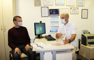
Today you do not need to go anywhere to establish a diagnosis. It is enough to apply to LISOD.
Success in the fight against cancer directly depends on the quality of diagnosis. The accuracy of the results guarantees the correct adequate treatment in the future. In LISOD, just like in the leading medical centers of the world, professionals and modern equipment are concentrated.
Today, there is no need to go abroad to undergo a quality examination. To establish an accurate diagnosis, you need to contact LISOD:
– patients are examined using the latest equipment, receive the conclusion of foreign experts;
– Israeli clinical oncologists prescribe only the necessary diagnostic procedures and manage the entire treatment process.
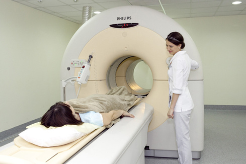
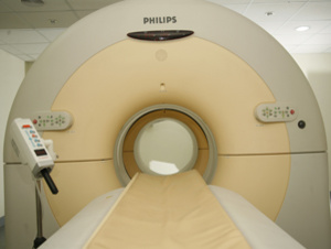
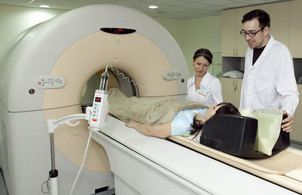
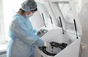
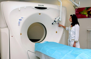
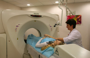
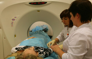
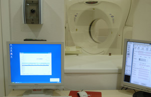
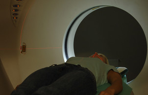
LISOD operates PET-CT. Any person, if necessary, can be examined on a positron emission tomograph. In the USA, in Israel, in the developed countries of Europe, PET-CT as a "gold" standard is used to diagnose 65% of oncological diseases: lymphomas, tumors of the stomach, intestines, mammary glands, head and neck, lung, prostate, pancreas, etc. Positron emission tomography is an innovative diagnostic method. PET-CT examination allows to detect a malignant tumor at the earliest stage of development, when the chances of successful treatment are high.
Being examined at LISOD means getting rid of uncertainty and starting treatment immediately. Patients have access to all types of modern diagnostics: mammography, computed tomography, diagnosis of diseases of the female genital area, etc. A highly qualified specialist performs endoscopic examinations (colonoscopy, gastroscopy, etc.), which are comfortable and painless for the patient. Pathological studies are carried out in leading certified laboratories in Germany and Israel, which ensures the accuracy of the diagnosis and, as a result, the correct treatment.
PET-CT RESEARCH
 Make an appointment
 Questions for LISOD
 Give feedback
PET-CT in questions and answers:
PET-CT is used to diagnose 65% of cancers. This is an innovative method that combines the capabilities of radiology and modern computer technology. The method is used as a standard in the developed countries of the world. Lung tumors, lymphomas, tumors of the head and neck, tumors of the stomach, intestines, tumors of the mammary glands (in many cases), tumors of the prostate, pancreas and others cannot be treated today without PET.
According to the European Association for Nuclear Medicine, one PET-CT unit per 1.5-2 million people is needed to ensure accurate early cancer diagnosis. For comparison: in Germany – 80 devices per 82 million people; in Israel – 6 PETs per 8 million population; in France – 45 PET machines per 65 million people. Thus, in order to provide assistance to cancer patients at the European level, at least 20 PET-CT facilities should operate in Ukraine.
Until recently, the only positron emission tomograph operated in the country. Many people who need a PET-CT examination due to indications did not have the opportunity to undergo it. Today, the situation has changed radically: a positron emission tomography device combined with computed tomography operates at LISOD. Each patient, if necessary, can undergo a PET-CT examination in order to receive adequate treatment and successfully overcome the disease.
PET-CT in questions and answers:
- PET-CT 24 600 UAH.
- Intravenous contrast for PET-CT (up to 6 hours) UAH 2,600.
- Oral contrasting for PET-CT 650 UAH.
Sign up for diagnostics
Or call +38 (044) 520–94–00
daily from 8:00 to 20:00
PET-CT is a diagnostic method and a method for monitoring the development of a neoplasm
Thanks to the use of PET-CT in LISOD, accurate diagnosis is now available to patients and, as a result, correct treatment is guaranteed. Based on the results of the examination, radiologists identify the location of the tumor and determine whether it is benign or malignant; specialists distinguish a neoplasm from an inflammatory process and establish, in the case of cancer, the prevalence of the tumor.
- PET-CT 24 600 UAH
- Intravenous contrast for PET-CT (up to 6 hours) 2 600 UAH
- Oral Contrast for PET-CT 650 UAH
Sign up for diagnostics
Or call +38 (044) 520–94–00 daily from 8:00 to 20:00
With the help of PET-CT examination, doctors find out the condition of the patient's tissues. The use of a positron emission tomograph makes it possible to determine cancer in the initial stage, at which there are still no significant changes in the tissues, and CT establishes the exact position of disease-affected cells. One study is being conducted instead of several. Israeli oncologists develop treatment tactics and make the necessary appointments, taking into account the specific characteristics of the patient's tumor.
In most cases, the use of a positron emission tomograph (PET-CT) further excludes a series of tests: thus, time is saved, which is vital in oncology, and human resources (after all, each additional study only increases the cost of treatment). The PET-CT examination procedure for a LISOD patient is a complete diagnostic procedure with interpretation of the results by a nuclear medicine doctor and radiologists trained in leading Israeli clinics.
The main advantages of the method:
- high accuracy;
- recognition of the disease at the earliest stage, which can not be done only with the help of CT, MRI and laboratory tests;
- Proper diagnosis.
PET as the newest method for assessing the effectiveness of treatment
In the process of treatment, after several cycles, it is possible to conduct a study again to compare new results and initial. This approach in many cases eliminates the need to repetition analyzes of neoplasms or further continuing treatment. For example, today adopted a standard for lymphoma: conducting a control PET after several chemotherapy courses; If there are no improvements, change treatment and achieve, thus maximum efficiency.
Using PET-CT for tumor irradiation
With the help of magnetic resonance tomography (MRI) and computed tomography (CT), images of tumor neoplasms are obtained. But not every formation contains cancer cells. Thanks to the use of PET, it is possible to significantly reduce the exposure zone and accurately carry out radiotherapy. In such cases, it is especially important for the patient that healthy tissues and organs suffer, the radiation load is reduced; As a result, there are no pronounced side effects.
PET CT Research, Photo Gallery


PET CT studies for diseases:
- Mammary cancer
- Brain cancer
- Cancer Gortani.
- Cancer Guba
- Gallbladder cancer
- Skin cancer
- Lungs' cancer
- Lymphoma
- Uterine cancer
- Melanoma
- Cancer Almonds
- Bladder cancer
- Liver cancer
- Esophageal carcinoma
- Cancer penis
- Crack Cancer
- Intestinal cancer. Rectal cancer.
- Cancer anal canal
- Cancer oral cavity
- Cancer bones
- Sarcoma Caposhi
- Cervical cancer
- Thyroid cancer
- Egg cancer
- Ovarian cancer
- Cancer Language
- Myelodsplastic syndrome
- Chronic lympholecosis
Endoscopic ultrasound
- Make an appointment
- Questions to Lisod.
- Give feedback
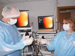
In Lisod, one of the newest methods for diagnosing diseases of the digestive system is widely used – endoscopic ultrasound or endusy.
An endosi uses a special endoscope equipped with an optical instrument and an ultrasonic sensor. Ultrasound penetrates deep into fabric, accurately transfers information that is transformed into a high quality image. The result is displayed on the monitor.
The most important factor for patients: endoscopic ultrasound has all the advantages of ultrasonic and endoscopic techniques.
The method allows you to reproduce internal organs in such small details that are not visible in the usual ultrasound. At the same time, the endusion has the advantages of the endoscope, i.e. the doctor when conducting a survey sees the entire inner surface of the digestive tract.
Endoscopic ultrasound is used to diagnose the following diseases:
- Colon cancer, duodenal cancer, stomach cancer, esophagus cancer;
- subliminate neoplasms of the stomach, esophagus, colon (lipoma, fibroma, leyomioma);
- ulcerative disease of the esophagus, stomach, colon and duodenal estate;
- varicose veins of the esophagus;
- Diseases of biliary tract.
In addition, enduri is used to diagnose and obtain material from formations that were previously not available for minimally invasive methods: tumors and mediastinal lymph nodes, liver gates, pancreas, small pelvis.
With the help of enduri, experts determine the scale of the spread of cancer (on lymph nodes and other organs), conduct biopsy to diagnose the state of the organs under study. Thus, doctors establish the stage of the disease and develop the most effective treatment plan.
Important. In Lisod, endusy is performed absolutely painless for the patient. Apply t. N. "Small" anesthesia: the patient falls asleep and does not feel anything; Awakening easy, without side effects. At the same time, the doctor has the ability to carefully conduct a study.
PET CT studies for diseases:
- Cancer of the subtle intestine
- Stomach cancer
- Esophageal carcinoma
- Pancreas cancer
- Prostate cancer
- Intestinal cancer. Rectal cancer.
Endoscopic research
- Make an appointment
- Questions to Lisod.
- Give feedback
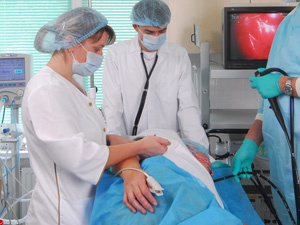
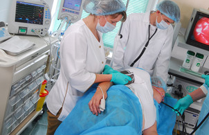
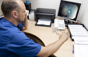
With very accurate digital video endoscopic equipment in Lisod, more than 20 endoscopic procedures are carried out:
- Colonoscopy (colonoscopy is a study of the walls of the colon);
- bronchoscopy (method of assessing the lumen of the bronchi, trachea and mucous membrane; use a flexible endoscope, which is introduced into the lumen of the trachea and bronchi);
- Visual phyagolaryngoscopy (study of the upper respiratory tract);
- retrograde cholangiopancratography (study of pancreatic and bile ducts of the pancreas);
- Ezophagogastroduodenoscopy (Gastroscopy – study of the gastrointestinal tract);
- stenting (special procedure to restore the normal state of the ducts); We carry out stenting the output department of the stomach, esophagus, colon, pancreatic duct, biliary ducts.
- chromoendoscopy (use of additional staining);
- Cistoscopy: With the help of a cystoscope (special tool), an optical examination of the bladder is carried out; Cistoscopy is a basic study for the diagnosis of urinary bubble diseases, urethra, kidneys;
- biopsy;
- percutaneous endoscopic gastrostomy;
- Endoscopic polypectomy – removal of polyps;
- Treatment of varicose veins of the veins of the esophagus and stomach;
- Endoscopic excision (resection) of the mucous membrane;
- Argonoplasma coagulation of the esophagus Barrett;
- Endoscopic dialation (expansion) of narrowings;
- Endoscopic Clipering (seam imposition);
- removal of foreign objects;
- Endoscopic bleeding stop (endoscopic hemostasis).
You need to know: during colonoscopy, simultaneously delete Revealed polyps, That is colorectal cancer prevention. Anyone can undergo a safe and effective colonoscopy in Kyiv in our hospital. As a routine screening procedure, we recommend a colonoscopy for all women and men over 50 years of age.
High image quality allows the doctor to detect even the smallest deviations from the norm. Painlessness and comfort during manipulations are of great importance for the patient.
Advantages of the method for LISOD patients:
- the procedures are absolutely painless: “minor” anesthesia is a short medical sleep (the patient falls asleep and does not feel anything); easy awakening without any side effects;
- research is performed by a highly qualified specialist;
- the use of modern equipment and the professionalism of doctors guarantee patients the accuracy of research results; the results are written to disk.
Important. The endoscopy department is equipped with the EVIS EXERA III CV-190 system from OLYMPUS. This is one of the world's best, rare for Ukraine, video information centers, which has a number of undeniable additional benefits for patients.
- A special technology of optical imaging allows examination of the mucous membrane without the introduction of a contrast agent. Thus, the number of manipulations is reduced and the time of the procedure is reduced.
- The 150x magnification allows the doctor to perform a thorough examination and see the state of the tissue in a narrow-spectrum mode – much more than with conventional chromoendoscopy. In particular, during a colonoscopy, a specialist already at the stage of the study determines whether changes in the vascular pattern of polyps correspond to malignant ones, or, conversely, do not carry the risk of degeneration.
Endoscopy, photo gallery
BREAST DISEASES
- Make an appointment
- Questions for LISOD
- Give feedback
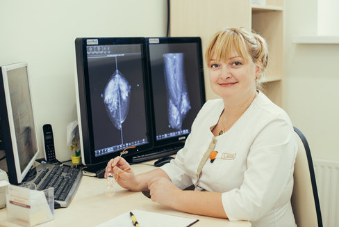
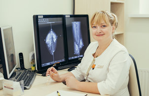
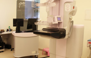
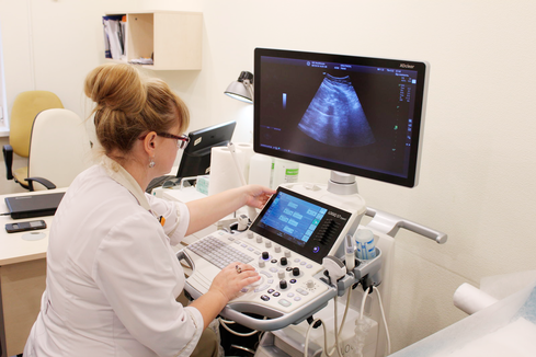
LISOD has an effective system for diagnosing benign and malignant diseases of the mammary glands. Depending on the age of the woman, an ultrasound or mammogram is performed. Examinations at LISOD are carried out quickly, comfortably and safely.
Ultrasound examination takes place on the most modern ultrasound diagnostic apparatus, which is equipped with a special sensor with a “doppler” mode. If necessary, 3D visualization is used to obtain more reliable information about a possible formation.
If a woman is older than 35 years, mammography is recommended, which is recognized as the “gold standard” for detecting asymptomatic and non-palpable tumors. If breast cancer has been diagnosed in close relatives, it is possible to have a mammogram at a younger age.
LISOD is equipped with the most modern digital mammograph with Giotto Class tomosynthesis. A huge advantage of 3D mammography is the ability to examine the mammary gland section by section and identify the slightest deviations from the norm.Today, such a diagnosis is the shortest way to the correct diagnosis. You should not be afraid of the negative impact of X-ray radiation: special filters are built into the device that protect the health of a woman.
Benefits of breast examination at LISOD:
- painless procedures;
- modern equipment provides not only information content, but also safety;
- specialists who have completed training in the leading medical centers of the world, with many years of experience in diagnosing breast diseases;
- obtaining a second opinion of a foreign specialist (if necessary).
LISOD performs all types of breast tumor biopsies: aspiration biopsy for cytological examination; stereotactic biopsy – for histological examination; ductography – the ducts of the mammary gland are examined; trucate biopsy – for histological examination.
The main purpose of the procedures is to determine the benignity or malignancy of the process.
The LISOD high-tech stereotactic biopsy. A special installation allows you to very accurately and painlessly take for analysis, and often completely remove a formation up to 1.5 cm in diameter.
Important. Vacuum aspiration of neoplasms is an alternative to surgical intervention.
Benefits of Lesions Removal in LISOD:
- "cosmetic" and minimally invasive – the procedure is carried out through a point incision;
- painless – performed under local anesthesia;
- absence of deformation and scars on the mammary gland, which simplifies the subsequent diagnosis;
- the possibility of histological analysis and complete removal of small benign formations.
All manipulations are performed with anesthesia on an outpatient basis. Procedures do not require special training. The materials obtained during the biopsy are sent to certified laboratories in Germany and Israel. This is essential for every patient, since the choice of the correct treatment plan depends on the results of histological studies.
Breast diseases, photo gallery
DISEASES OF THE URINARY SYSTEM
- Make an appointment
- Questions for LISOD
- Give feedback
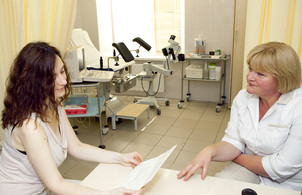
Diagnosis of diseases of the genitourinary system
The Center for Modern Urology LISOD has all the technical capabilities to conduct a comprehensive examination of patients. This allows you to quickly and accurately establish a diagnosis, prescribe effective treatment.
Important! We employ experienced urologists with many years of experience in diagnosing and treating inflammatory diseases, benign and malignant neoplasms of the genitourinary system. Doctors act in accordance with international medical protocols and never prescribe unnecessary examinations.
At the consultation appointment, the doctor necessarily examines the patient. Taking into account complaints, personal and family history, clinical examination, diagnostic methods are determined to identify the disease.
Methods used to diagnose urological diseases:
- Clinical blood tests.
- Laboratory studies of urine, urogenital smears, the proportion of the prostate gland.
- If necessary, men are analyzed on a monacarker – a prostate-specific PSA antigen.
- Ultrasound examination is used for the primary diagnosis of many diseases. Men are underway urinary tract (kidneys, bladder), prostate, testicles, transrectal prostate ultrasound. Women are performed ugly urinary tract (kidney, bladder).
- Cistoscopy: With the help of a cystoscope, an optical examination of the bladder is carried out (used to diagnose urinary bubble diseases, urethra).
- Ureteroscopy: With the help of a ureteroscope, an optical overview of the ureter and a cup of kidney system is carried out (used to diagnose uretera and kidney diseases).
- X-ray-contrast methods (in particular, overview urography, excretory urography, etc.).
- CT scan.
- PET CT.
- Biopsy for the implementation of histological examination (in particular, multi-flow biopsy of the prostate gland, the puncture biopsy of the kidney under UZ-control).
- Immunohistochemical studies.
- Molecular studies.
Urological diseases that are diagnosed and treated in Lisod:
- benign and malignant neoplasms of kidneys, ureters, bladder, prostate gland, eggs, penis;
- acute and chronic cystitis;
- acute and chronic prostatitis;
- prostate hyperplasia;
- acute and chronic pyelonephritis;
- Balanopostitis;
- epididimitis;
- kidney cysts;
- Urolithiasis and others.
Advantages for Patients of the Center for Modern Urology Lisod:
- High class specialists.
- Maximum decreases time from the patient's appeal to consult before establishing the diagnosis and treatment of treatment.
- Comfortable reception conditions are provided, attentive attitude of medical personnel.
- The most modern approaches are applied, specialists act in accordance with the recommendations of the world's leading urological societies.
- Professionalism of doctors and the use of modern equipment guarantees the accuracy of research results.
- Preventive inspections and surveys are carried out. This allows you to make sure that everything is in order with the body or identify dangerous diseases at an early stage, when there are no symptoms and at the same time the highest chances to fully cure the disease.
Remember! Appeal to a qualified doctor and timely diagnostics allow you to get the best results of treatment. Do not delay a visit to a specialist if there are incomprehensible symptoms.
Diseases of the urogenital system, photo gallery
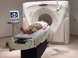
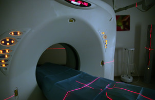
CT SCAN
- Make an appointment
- Questions to Lisod.
- Give feedback
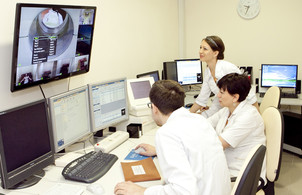
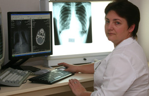
The department is carried out with computed tomography (CT) of the head and neck, upper and lower departments of the abdominal cavity, pelvis, joints, spine, limbs. CT Urography is also carried out – x-ray study of the urinary system organs to assess the excretory function of the kidneys and changes in the urinary system and the surrounding tissues. For patients, it is an important role to monitor the results of all diagnostic CT research by the Lisod radiologists who have been internship in the leading medical centers of Israel.
Advantages of the method for Lisod patients:
- The use of multi-section computer tomograph made by Philips;
- Contrasting to improve visualization (it is very important for oncological patients: artificially changes the density of tissues, and the visibility of individual structures is enhanced);
- Conducting CT-urgent;
- Performing all types of biopsies (soft fabrics, liver, bones, light, mediastinal organs) under the control of the CT to install an accurate diagnosis.
Computer Tomography, Photo Gallery
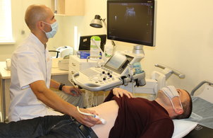
Ultrasound
- Make an appointment
- Questions to Lisod.
- Give feedback
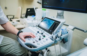
In Lisod, ultrasound diagnostic equipment is equipped with various types of sensors, which allows ultrasound research: abdominal cavity, small pelvis, transvaginal, transrectal, kidneys and bladder, breast, thyroid gland, neck, testicles, hearts, soft tissues.
Control of ultrasound perform puncture and other manipulations (for example, puncture of the prostate gland, the thyroid gland, pleural and lapropuncture, the production of nephrost, etc.).
Advantages of the method for Lisod patients:
- use of special equipment of the expert class;
- lack of radiation load on the patient;
- Equipment of various types of sensors for research;
- Research performs a highly qualified specialist.

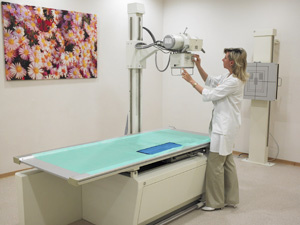
X-ray diagnostics
- Make an appointment
- Questions to Lisod.
- Give feedback
In the department, important radiological studies of the bodies of the chest cavity, the abdominal organs and the pelvis, the bone-articular system are carried out. With the help of special equipment, X-ray control of many invasive surgical procedures are performed.
Advantages of the method for Lisod patients:
- Modern and safe equipment;
- A wide range of procedures: X-ray studies of breastfeeding organs, abdominal organs and pelvis, bone-artistic system;
- Execution of x-ray diagnostics by lying patients (there is a mobile x-ray).
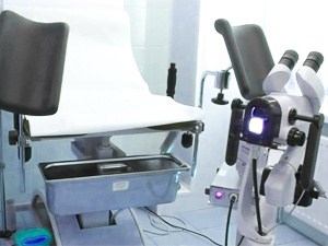
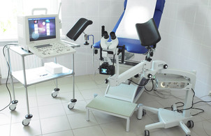
Oncogynecology
- Make an appointment
- Questions to Lisod.
- Give feedback
Oncogynecology is at the junction of gynecology, oncology, urology, endocrinology. It is to the oncogynecology that refers to the management of diseases such as cervical cancer, ovarian cancer, uterus cancer, vaginal cancer, vulva cancer.
Each woman should know: only a professional can identify a disease or confirm its absence.In the department of oncogynecology, Lisod is diagnosed and the treatment of all diseases of the female sexual sphere: both benign and malignant.
Oncogynecology in Lisod – Benefits for Patients:
- Acceptance of the highest qualifications: Professor, Doctor of Medical Sciences, World Specialist;
- Comprehensive examination includes colposcopy, vaginal ultrasound examination, cytological studies, various types of biopsies, etc.;
- Due to the qualitative diagnosis, the consultation of specialists approves the only faithful treatment plan.
Extremely important for woman Prevention of cervical cancer. In Lisod spend:
- diagnosis and timely treatment of precancerous diseases;
- Vaccination against papillomavirus infection, warning cervical cancer.

Laboratory of Patomorphology

- Make an appointment
- Questions to Lisod.
- Give feedback
Patomorphological verification of diagnosis – the basis of diagnosis and treatment in modern oncology. The most important advantage for Lisod patients: Research is carried out in leading pathological laboratories of Germany, Israel and Greece. In the partner laboratories, all samples of tissues obtained when performing operational interventions and biopsies are sent; Cytological and histological preparations are also sent.
It should be understood that the erroneous result of research can be a fatal for the patient: treatment may be lazy without needing or not begun on time in case of acute necessity, incorrect treatment can be carried out. That's why The cooperation of Lisod with foreign certified laboratories is of great importance.: According to the results of research, doctors confirm with absolute reliability or refute the oncological diagnosis and, in case of cancer detection, the only faithful treatment plan is produced.
Additional research is performed in the hospital: PCR, PT-PCR, Fish-Research On fabric. For patients, it is important that, according to the results of these studies, the testimony is determined to carry out various types of chemotherapy, including targeting (targeted) therapy.
In Lisod use Modern methods of molecular and genetic research Patient blood and cancer cells. As a result of these studies, specialists establish whether the patient has a predisposition to the occurrence of cancer; In the case of oncology, the degree of aggressiveness of the malignant tumor is determined and the optimal treatment plan is developing.
What advantages give patients molecular research?
- The result may show that in some cases the treatment will be limited only by surgery: even without chemotherapy and radiotherapy will not be refunded.
- An analysis of some cancer cell growth receptors is performed to block them with special antibodies and, thus, to prevent their reproduction in the future.
- Genetic changes in tumor cells are determined, which makes it possible to find out whether the formation is amenable to targeted therapy or some type of chemotherapy.
LISOD specialists recommend the molecular test for melanoma, ovarian cancer, breast cancer, head and neck cancer, pancreatic cancer, and lung cancer. Molecular analysis will help many patients who have already undergone standard treatment; will also help patients with rare cancers and patients with metastases.
In LISOD, intraoperative (immediately during the operation) pathomorphological studies are performed, which play an important role for the patient: during the operation itself, doctors decide on the extent of the surgical intervention.