Bullae in the lungs (emphysema) Bullae (false pulmonary cysts) are pathological air cavities in the lungs (from the English blebs – “bubbles”) that can occur due to mechanical damage
Bullae in the lungs (emphysema)
Bullae (false pulmonary cysts) are pathological air cavities in the lungs (from the English blebs – “bubbles”) that can occur due to mechanical damage to the parenchyma, an infectious-inflammatory or other disease. Excess air in these sac-like cavities, changes in the structure of the lung matrix, and a reduction in the area of functional areas of the respiratory organ lead to persistent respiratory failure, and the consequences of such pathological changes can be irreversible. The formation and enlargement of bullae in the lungs leads to a decrease in the gas exchange function of the lungs, and in the event of a rupture of a large bulla, it can lead to a life-threatening condition – pneumothorax.
An interesting fact: for patients with large bullae in the lungs, air travel is dangerous. The fact is that in flight at an altitude of 2.5 km from the ground, pathological cavities can increase in diameter by 30-40%, which means that an episode of pneumothorax, bulla rupture, can occur.
Bullae are most often diagnosed in people who smoke (usually men over 50 years of age with about 15 packs of experience) against the background of centrilobular emphysema or COPD. Both of these diseases are somehow associated with damage (stretching) of the alveoli, a violation of the structure of the lung parenchyma, with its aging. Sometimes these pathological changes are not associated with smoking, but are the result of a metabolic disorder, an infectious-inflammatory process, an autoimmune disease, or are genetically determined.
The disease is difficult to treat. If the cause of the formation of bullae is established, their size is small, diagnostic measures are taken on time and treatment is started, pathological changes are amenable to conservative treatment. In pneumothorax, the bullae are removed surgically using endoscopic techniques.
Bullae require dynamic monitoring: CT scan, spirometry, consultation with a pulmonologist.
What does bulla mean in the lung?
According to the definition given at the international medical CIBA symposium, a bulla should be considered an air cavity in the lung, the diameter of which exceeds 1 cm, while the alveolar wall becomes thinner to 1 mm or less.

In medicine, it is customary to distinguish between bullous emphysema and bullous disease. They are distinguished depending on the causes of occurrence, according to etiology, and also depending on the consequences for health. Bullous emphysema is usually diagnosed in patients with signs of COPD (chronic obstructive pulmonary disease) on the background of bilateral panacinar or centrilobular emphysema. With bullous disease, emphysema is not observed, and the cavity may be one or slightly larger.Bullese disease can be with congenital marfana syndrome. Bullese emphysema in the absence of treatment and adequate actions to contain its growth (refusal of smoking, working on hazardous production, treatment of chronic bronchitis, etc.) with time can lead to respiratory or heart failure, a collapse of the lung, heavy coughing with blood, to intolerance to physical Loads.
Bullys are formed on the principle of soap bubbles: first there are small, then they are combined, forming large bulls, and so on. With a bullous emphysema, the pulmonary cloth becomes similar to the old sponge – as it is wearing the pores, becomes larger, and the material loses elasticity and breaks.
Large bullies (diameter can reach up to 10 cm) are formed during the destruction of alveolar partitions. Alveola – bubbles, of which there is a normal air pulmonary fabric. Air storage, oxygen transportation, air exchange is carried out in alveoli, and the process itself requires the structure of the dosage intake. Bully, even very small, bring the element of chaos and entropy into the work of the most important respiratory body.
Symptoms of bullous emphysema
In a number of studies published in the open international base PubMed, it is emphasized that the formation of Bull in the lungs inevitably cause such factors as:
- Smoking (15-20 Pachcolat);
- Systematic inhalation of toxic substances (unfavorable ecology, work in harmful production, toxicization, drug addiction);
- Low level of quality of life (scarce and harmful meals, supercooling, disease, lack of qualified medical care).
And only after these factors in the prevalence, infectious inflammatory diseases of the respiratory tract, autoimmune diseases, disruption of metabolism, congenital characteristics of the body are followed by the prevalence.
In most cases, the bullous emphysemia is diagnosed in people of old or mature age in people, however, Bullas can be found in young people.
Small bulls do not show themselves. In general, most of the diseases of the main respiratory body patient is difficult to even suspect – the lungs do not hurt, and when shortness of breath, cough, lack of air, the disease leads the patient in resuscitation in a matter of hours.
Pathological symptoms can manifest themselves as the Bull and the development of emphysema increase. The prevention of the disease is possible with the help of regular screening – low-dose CT lungs. Examination in 3D mode and high resolution will show even the slightest deviations from the norm.

To determine the (accurate stage) of the bulwery emphysema, the method of spirometry is used – the functional diagnosis of the lungs. As a rule, after spirometry, if the indicators are not normal, the pulmonologist sends the patient to the PT of the lungs.
To suspect the bully in the lungs in the presence of the following symptoms:
- Shortness of breath and respiratory impairment;
- Physical exertion resistance, fast fatigue;
- Hypotension uncontrolled arrhythmias;
- Feeling of constant fatigue;
- Singing lips and fingertips;
- Painful pallor;
- Feeling of incompication;
- The deformation of the chest (becomes a mustache, if there are large bulls).
Since the increase in Bull is quite a long time in time, the patient may not notice deterioration of well-being, in particular, it becomes more and more difficult to delay the breath due to the decrease in the life capacity of the lungs, and hypoxia affects all internal organs – a person literally "fades".
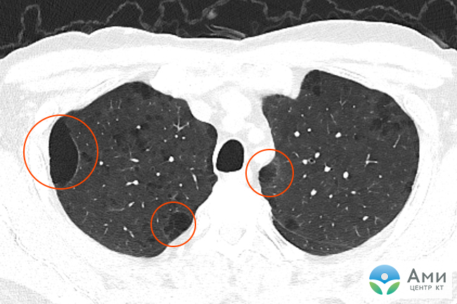
Bully in the lungs on CT
Bullys in the lungs on CT are visualized as distinct, relatively more darkened bubble sections of lung tissue, reminiscent of holes or large pores of the sponge. The more dense fabric, the more it is bright on the CT-scannes – therefore, for example, the bones are white, and the air pulmonary fabric is relatively homogeneous graphite-gray.
An X-ray rhythmologist can accurately determine the diameter of Bull, their quantity, to find out if there are signs of emphysema, bronchiectases or other diffuse pulmonary diseases.
How to treat bully in the lungs
The patient needs to abandon smoking and leave work in harmful production activities, otherwise all therapeutic activities will be meaningless. In uncomplicated cases and in the early stages of emphysema (or bullous disease), treatment conservative, combined. Therapy is appointed by a pulmonologist and may include:
- Medicines (diuretics, bronchopholics, hormonal drugs);
- Special course of exercises and respiratory gymnastics;
- Regular oxygen therapy with the use of an oxygen concentrator.
- Regular diagnostic measures (CT lungs, spirometry, consultation of the pulmonologist).
A positive aspect is the fact that the full and lifelong refusal of smoking in most patients suspends the development of bullous emphysema, and the use of oxygen therapy in serious patients can extend life for 5-10 years.
Surgical treatment of Bull (endoscopic "stitching") can be shown in recurrent pneumothoraxes against the background of growing cavities. The patient is carried out bullctomy and Pulberry operations. However, the absolute success of such treatment cannot be guaranteed.

Surgery
Surgical treatment is called bullctomy. If a person has been revealed by a giant Bulla – this is an indication of surgical treatment. Small dimensions of Bull are poorly sufficiently surgical treatment, the effect of the operation is insignificant. Dyspnea rarely decreases.
Bully, not accompanied by clinical symptoms (shortness of breath) or complications (pneumothorax, suppuration), is not recommended.
Contraindications to bullectomy are continued smoking, severe cardiovascular disease, diffuse emphysema with little compression of the surrounding lung tissue.
The indication for surgical treatment may be recurrent pneumothorax against the background of bullous changes. In this case, bullectomy and pleurodesis are performed simultaneously.