Detailed description, symptoms, methods of treating and preventing breasts of the knee, elbow and shoulder joints. The reasons for the appearance of bursita.
Bursit – reasons and effective types of treatment of bursita
Bursit (bursa) translated from Latin – bag.
Bursitis is an inflammatory disease of the synovial bags, accompanied by increased education and accumulation in their cavities of exudate.
The company ESMA represents professional medical equipment and apparatuses for the treatment of bursita and other diseases and injuries.
Equipment for sports recovery medicine.
Clinical flow bursts can be sharp, subacute, chronic, recurrent; According to the nature of the pathogen – nonspecific or specific (gonorial, brucellular, tuberculosis, syphilitic); According to the nature of the exudate – serous, purulent or hemorrhagic. Some authors allocate aseptic and infected bursitis.
Sinovial bags – Sugal cavities formed by the synovial shell containing the synovial liquid. Sinovial bags belong to the auxiliary apparatus of muscles. They are located in the fiber between the protruding sections of bones and soft tissues (skin, fascia, muscle tendons, muscles). The following synovial bags are distinguished: subcutaneous – located in the subcutaneous tissue on the convex surface of the joint exposed to sharp bending; subfascial – under fascia; Docking – located under the tendons; Mortal – lying under the muscles. Docking synovial bags in adults are often communicated with the joint cavity. Surrounding the muscle tendon at a considerable mass, they can form a synovial tendon vagina.
EXUDAT. – liquid rich in protein and containing uniform elements of blood; It is formed when inflammation.
The inflammatory process may occur both in constant and in new-formed bags in places of continuous pressure and friction of the skin, fascia, muscles, tendons about bone protrusions. Most often occurs to bursitis the elbow synovial bags and the synovial bags of the knee and shoulder joints. The frequency of such localization is due to constant injury to the corresponding parts of the body due to the peculiarities of certain types of sports. This makes it possible to take a bursitis towards professional diseases. The frequency of morbidity bourcitis depends on the sport and other variables (sporting regimes, equipment, the level of skill, etc.).
Causes of Bursita
The reason for the occurrence of acute bursitis is more likely to be injured – injury, abrasion, small wounds and secondary infection of the synovial bag with gloty microbes. The infection of synovial bags occurs according to the lymphatic paths from purulent foci (in the face of inflammation, boils, carbuncules, osteomyelitis, proleells), and infection through blood is not excluded.Also, the possibility of infection is not excluded due to cut or abrasion in the articular bag area (when you fall from a bicycle, when playing football). Chronic bursitis is often a consequence of a long constant mechanical irritation. Pathological anatomy. Pathoanatomic changes in acute bursite are expressed by signs of acute inflammation of the walls of the synovial bag.
The initial stages of acute bursitis are characterized by serous impregnation of tissues and accumulation in the cavity of the serous exudate bags (acute serous bursitis). If there is a microbial flora, serous inflammation is quickly quickly moving into purulent (purulent bursitis). The spread of the purulent process to the surrounding tissues can flow according to the type of phlegmous inflammation with the necrosis of the wall of the bag and the formation of subcutaneous and intertensular phlegm. In the launched cases, no longer healing fistulas are formed. The breakthrough of the pus in the cavity of the joint leads to the development of purulent arthritis.
With acute traumatic rusty in the stretched synovial bags and their pockets accumulates hemorrhagic fluid (blood or plasma). With reverse development, the organization of fibrin and obliteration of the blood vessels of the synovial shell occurs. Resistant changes in the wall of the bag are developing, which thickens, the surface of the synovial shell is covered by the expansions of the connective tissue (proliferating bursitis), dividing the cavity of the bag for additional pockets.
When the acute inflammation is subsided and at a subacute of the flow of rusty in the wall and pockets of the bags, there are encapsulated sections of necrotic tissues or exudate, which, during repeated injury and infection, serve as a favorable soil for the development of inflammation recurrence (recycling bursitis).
Symptoms of Bursita
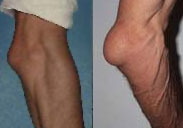 At the site of the anatomical arrangement of the bag, the rounded resistant painful swelling of soft-elastic consistency is determined, fluctuating. The diameter of the swelling can reach 8-10 cm (Fig. 1). The patient complains of pain in the field of swelling, malaise; Increases body temperature; The joint function is moderately limited.
In phlegmonous inflammation, there is no swelling of the tissue-surrounding bag, hyperemia of the skin (lymphangitis), are expressed (especially with gonorrheal bourrsites) common symptoms of the disease: severe pain, temperature rise to 39-40 °. When progressing inflammation and switching it to soft tissues, signs of phlegmon are determined.
With chronic roaring on the site of the bag, there is a rounded swelling of a soft consistency, the skin above it is moving, not changed, the function of the limb is not violated. Chronic process can exacerbate; In this case, the amount of fluid in the cavity of the bag increases, which sometimes leads to the formation of an isolated cystic cavity, filled with a liquid, called a hygroma.
Features of bursts of different location
Shoulder joint
In the area of the shoulder joint, bags that do not communicate with the cavity of the joint, subcutaneous acromial, adhesive and subcroids are most often affected (Fig. 2). Complaints are reduced to pains when the upper limb is allotted and rotating. Especially painfully rushes a faded bag. 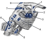
- Bag between the legs of a beak-acromic bundle
- Bag of the Cravoy-Shoulder Muscle
- Capsule of shoulder joint
- Bag of subband muscle
- Big Round Muscle Bag
- Bag of the widest muscles of the back
- Subakromial bag
- Acromic subcutaneous bag
In case of inspection, the shoulder contours are smoothed, there is a seemingly uniform increase in the delicid muscle itself; For large sizes of the bag, swelling is visible along the outer surface of the shoulder. Palpation is usually determined by soreness at a pressure on the inner edge of a large berry bone bulb. The per capita bursitis often has to differentiate from purulent arthritis, as well as from a very common disease of the shoulder joint – shoulder-blade periatritis.
Lock Susta
The acute bursitis of the elbow bags is more often the result of mechanical damage and infection of the subcutaneous elbow bag during its injury. The bag increases sharply in size and acquires a hemispherical form. 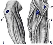 The addition of infection is accompanied by a sharp pain, redness of the skin in the area of the elbow process, increasing the temperature. A sharp bursitis is often striking the leaping bag located between the ray bone and tendon of the double-headed muscle (Fig. 3).
- Elbow subcutaneous bag
- Inter-site elbow bag
- White bag
Hip joint
Bursit in the area of the hip joint is distinguished by the severity of the flow; Inflammation often applies to the hip joint. It is more often inflamed by a deeply located ileal and tombechnaya bag, which is between the muscles and the articular capsule, as well as the surface and deep bags of a large spit. With purulent inflammation of these bags, there is a sharp pain when all the limb, extension and rotation of the thigh. The thigh is in the bending position, allotted and slightly turned outward. When palpation is determined by painful, elastic consistency swelling on the front-medial surface of the thigh under the groin bond. With purulent inflammation of the bags of a large spit, the swelling is more often located along the outer surface of the thigh. According to the severity of the clinical course and similarity of symptoms, these types of bursitis in some cases are difficult to distinguish from purulent inflammation of the hip joint. When bursites, unlike arthritis, are observed: 1) the relative painlessness of bending and bringing the thigh; 2) the absence of pain in the load on the length of the length; 3) The presence of swelling, located on the front-inner side of the hip below the groove bundle.To distinguish the defeat of the subcutaneous bag of the greater trochanter from the defeat of the deep bag allows the displacement of the latter during posterior movements of the thigh; with inflammation of the subcutaneous trochanteric bursa, no displacement is noted.
Knee-joint
In the area of the knee joint, the prepatellar bags are most often affected – subcutaneous, subfascial and subtendonal (Fig. 4); they do not communicate with the knee joint. Due to its more superficial location, the subcutaneous bursa is most commonly affected. Inflammation is accompanied by a sharp local edema, fluctuation, fever, and an increase in regional lymph nodes. Inflammation of the deep subpatellar bursa occurs more often secondary as a complication of acute persecution. Popliteal bursitis is difficult to diagnose due to the deep location of the bag; with it, there is some limitation of movement and pain in the knee joint.
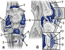
- 1-6. joint capsule
- Deep knee pouch
- Superficial pretibial bursa
- Semimembranosus sac
- Medial gastrocnemius bursa
- Upper patella bag
- Subcutaneous prepatellar bursa
- Subfascial prepatellar bursa
- Tendonal prepatellar bursa
Ankle joint
A common type of bursitis in the foot area is inflammation of a large bag located between the calcaneal tubercle and the calcaneal tendon – Achilles bursitis. It is caused by traumatization of the synovial bag with shoes, hematogenous or lymphogenous infection. Bursitis of the subcutaneous calcaneal bursa is a painful swelling in the region of the calcaneal tubercle. The inflammatory process may be limited to the phase of serous impregnation or lead to abscess formation. In some cases, this type of bursitis has to be differentiated from inflammation caused by soft tissue injury with a heel spur.
Diagnosis uncomplicated in inflammation of superficial bursae and is based on the typical clinical features described above. The diagnosis is facilitated by a puncture of the cavity of the bag, which makes it possible to establish the nature of the inflammation (serous, purulent, purulent-hemorrhagic, etc.) from the contents obtained, to establish the nature of the microbial flora and its sensitivity to antibiotics.
For the most effective treatment, it is important to exclude the specificity of the infection that caused inflammation (gonococci, brucella, spirochetes, etc.), which is possible on the basis of a carefully collected history, bacteriological examination of the contents of bags, and the results of specific serological reactions. The main differential diagnostic feature that distinguishes bursitis from arthritis is the preservation of movement in the joint. X-ray diagnostics of superficially located (subcutaneous) bursitis of any localization is relatively simple.Due to their availability for clinical recognition, it usually has only a clarifying character. X-ray diagnostics of deep bursitis is of much greater practical importance. Among them, the radiologist most often encounters bursitis of the knee joint and greater trochanter of the thigh, Achilles bursitis and inflammation of the inconsistent heel mucosa, and chronic subacromial bursitis on the upper limb.
In acute bursitis in the early stage, rest, pressure bandage, warm compresses are recommended. To prevent purulent bursitis, early active treatment of the serous form of acute bursitis, the use of fixing bandages is necessary.
In chronic bursitis, they often resort to a puncture with the removal of exudate and subsequent washing of the cavity of the bag with solutions of antiseptics or antibiotics. In case of traumatic bursitis, a hydrocortisone solution is injected into the cavity of the synovial sac (25-50 mg with antibiotics 2 to 5 times after a preliminary injection of 8-10 ml of a 2% novocaine solution). Careful observance of an asepsis is important, since serious complications are otherwise possible. With purulent bursitis, puncture treatment is used. In case of progression of the process, they resort to opening the bag and removing pus; a purulent wound is treated according to general rules. The disadvantage of this method is the duration of healing of the surgical wound.
Forecast in acute bursitis depends on the degree of pathological changes in the tissues of the affected bags, their prevalence, the ability of the infection to spread, the resistance of the patient's body. Unfavorable outcomes of acute bursitis can occur when it is complicated by arthritis, osteomyelitis, fistulas, sepsis. Recurrence in chronic traumatic bursitis occurs in 2-2.5% of operated patients.
Prevention consists in the elimination of permanent injury to the synovial bursae, in particular, wearing protective bandages, in the careful primary treatment of wounds of the synovial bursae with antiseptics (treatment with hydrogen peroxide, the application of a bactericidal patch / bandage), in the timely and rational treatment of pustular diseases.
Professional medical equipment and devices for sports rehabilitation medicine ESMA.
The photo shows the following models of devices:
Computer medical equipment – apparatus – complex ESMA 12.48 FAVORITE
Multifunctional portable device ESMA 12.04 MINIMAX