Computed Tomography (CT) – obtain information on this topic from the MSD MANUALS User Edition.
Computed tomography (CT)

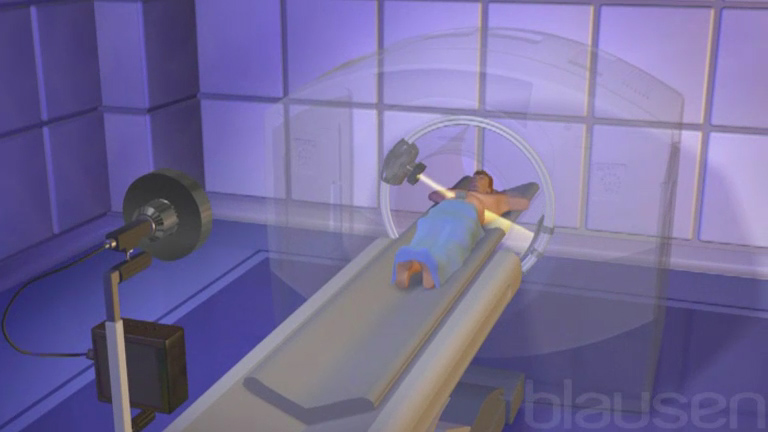
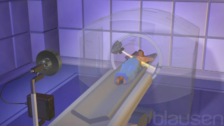
In computed tomography (CT), formerly computed axial tomography (CAT), an x-ray source and an x-ray detector rotate around the patient. In modern scanners, the x-ray detector usually has 4–64 or more rows of sensors that detect x-rays passing through the body. The data received from the sensors represents a series of x-ray measurements taken from different angles. However, the measurements are not immediately visible, they are sent to the computer. The computer converts them into images resembling two-dimensional slices (transverse). ("Tomo" in Greek means layer). The computer also creates 3D images from the captured images.
CT procedure
For a CT scan, the patient must lie on a movable table that moves through a donut-shaped opening. The patient is moved through the scanner and the devices rotate around the patient. For some CT examinations, the table moves periodically and stops with each image (slice) taken. In other CT examinations, the table moves constantly during the procedure. With helical CT, the patient moves in a straight line and the detectors move in a circle, so that a series of measurements is taken in a spiral around the patient – hence the term "spiral CT".
The patient's clothing should be free of metal buttons, snaps, zippers, or other metal objects in the area being examined, and all jewelry should be removed. These items are not dangerous, but they can block x-rays and distort the image. During the examination, the patient should lie still and periodically hold their breath when X-rays are taken to ensure that the image is clear. During the procedure, the patient may hear a buzzing sound.
Depending on the area of interest and scanner model, the procedure usually takes from a few seconds to several minutes. A chest CT scan takes less than a minute and patients only need to hold their breath once for a few seconds.
During CT scans, patients may be injected with a radioopaque contrast agent. Radiocontrast agents In imaging studies, contrast agents may be used to differentiate a single tissue or structure from its surroundings and to enhance detail. Read more. Contrast agents are the means that can be seen on the pictures, they help to distinguish one tissue from another.The contrast agent is injected into a vein, taken orally, or injected through the anus. The choice of contrast agent used depends on the type of examination and on the part of the body being examined.
The CT scan procedure is usually performed in an outpatient department. After the examination, patients can immediately return to their normal activities.
Visualization of internal structures: Computed tomography
In a CT scan, the scanner generates and captures x-rays by rotating around the patient, who moves through the scanner on a movable table. On one side of the scanner is an x-ray tube that generates x-rays, and on the other side is an x-ray detector.
Application of CT
High-resolution images provide more information about tissue density and the location of abnormalities than plain radiographs because doctors can pinpoint structures and abnormalities. CT allows the doctor to differentiate different tissues, such as muscle, fat, and connective tissue. Thus, a CT scan provides a clear image of organs not seen on plain radiographs and is best suited for visualizing most structures in the brain, head, neck, chest, and abdomen.
CT can detect and present data on any disorder in almost any part of the body. For example, doctors can use CT to identify a tumor, assess its size, pinpoint its location, and determine how far it has spread to nearby tissues. CT can also help doctors monitor the effectiveness of treatments (such as antibiotics for a brain abscess or radiation therapy for a tumor).
Types of CT
CT angiography
CT angiography uses CT and a radiopaque agent to create 2D and 3D images of blood vessels, including the arteries that supply blood to the heart (coronary arteries). their objects, as well as for finer detail. Read more. The contrast is injected into a vein (not an artery as in standard angiography), usually in the arm. Images are then rapidly obtained at such an interval that the radioopaque contrast can be visualized passing through the blood vessels under study. Using digital technology, a computer removes all tissue from the image except for blood vessels. (See also Coronary angiography Coronary angiography Cardiac catheterization and coronary angiography are minimally invasive examinations of the heart and blood vessels supplying the heart (coronary arteries) that do not require surgery. Read more  ).
).
CT angiography is used to identify:
narrowing or blockage (for example, blood clots) arteries;
protrusion (aneurysms) and breaks (bundles) of large arteries;
Anomalous blood vessels feeding blood to tumors.
CT angiography is usually used instead of standard angiography angiography when conducting angiography X-rays provide a high resolution of blood vessels. Sometimes this procedure is called standard angiography to distinguish. Read more information, since this is a safe and less invasive procedure (it is not required to install a catheter in the artery, which is associated with a slightly higher risk than when installing the catheter in Vienna). CT angiography visualizes the pathology of blood vessels is also exactly as magnetic resonant angiography, but slightly less exactly than standard angiography.

